Kunming, China, October 27, 2016
Osteoarthritis (OA) is a global degenerative joint disease that affects over 10% of the world’s population aged 60 years or older. Despite the recent availability of more advanced imaging modalities, controlled investigations of structural and biochemical changes in human osteoarthritic joints are still restricted by the relative inaccessibility of diseased tissues for sampling and the ethical limitations in the use of sham interventions or placebo, and in obtaining control human tissues. Thus, animal OA models are used to study the disease pathogenesis and to evaluate the potential effects of various therapeutic agents intended for human/clinical use.
Spontaneous OA occurs in nonhuman primates and is known to affect over 65% of cynomolgus monkeys >15 years of age, and about 30% of cynos between 10-15 years old. The similarity between human and NHP OA in terms of age-related prevalence, clinical course and disease severity and, sequential anatomical and biomechanical degenerative changes make the cynomolgus macaque an excellent translational disease model for human OA. This model can be particularly useful to evaluate the pharmacodynamic action and therapeutic efficacy of novel pharmaceutical entities. Furthermore, the availability of behavioral, functional and activity assessment pain measures in the monkey disease model allows more effective characterization and investigation of OA pain response and modulation patterns following the administration of prospective pharmacological therapies that are intended for human/clinical use.
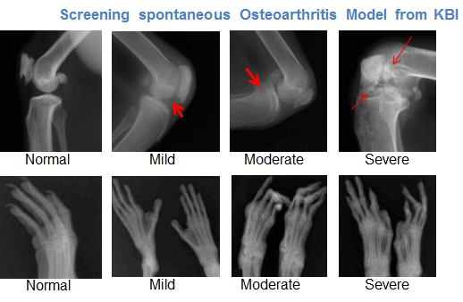
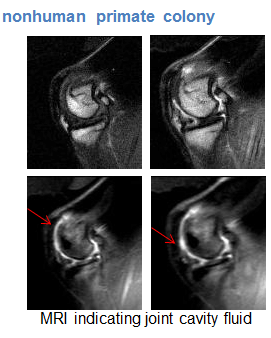
X-ray of the joints can be reliably performed at KBI for the screening of naturalistic arthritis models from KBI’s large population of aged cynomolgus monkeys. Typical x-ray changes characteristic of OA such as joint space narrowing, subchondral sclerosis, subchondral cyst formation, and osteophytes have been observed in our naturalistic cyno OA disease models. Furthermore, magnetic resonance imaging (MRI) can effectively be utilized at KBI for the longitudinal assessment of semi-quantitative and quantitative imaging endpoints, such as cartilage morphology and composition, of the affected joints and articular tissues.


Contact us if you need more information.